Four-dimensional computed tomography (4D-CT) holds promise in enhancing the comprehensiveness of pulmonary scans but is often limited by the scanning capacity. Conventional 4D-CT scans cover only 16 cm along the axial direction z-axis with 320 slices, insufficient to capture the entire lung in a single session. This study aims to construct a complete “full-lung” volume by synthesizing scans from different segments of the lung at matching respiratory phases, with an emphasis on matching vascular structures and other critical anatomical features.
4D-CT imaging stitching
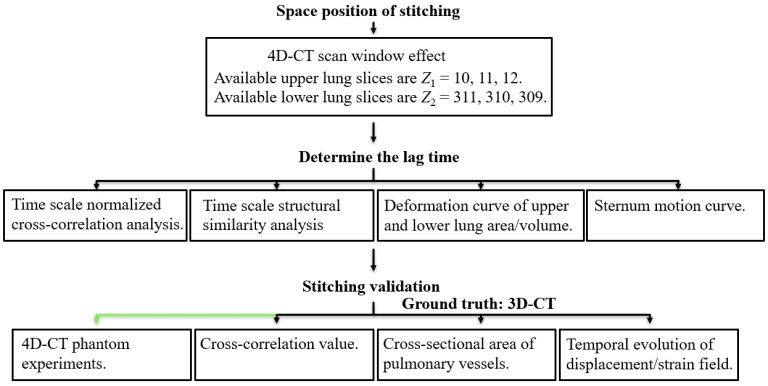
The upper and lower sections of the lung were scanned separately during controlled breathing cycles. The scans were synchronized temporally and spatially using approaches based on the image cross-correlation algorithm, deformation of the lungs, and sternum displacement, allowing for the accurate stitching of voxel data from identical respiratory phases.
Fig. 1. Stitching algorithm and validation map.
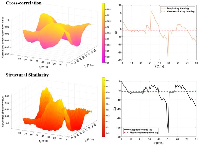
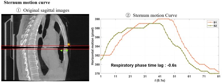
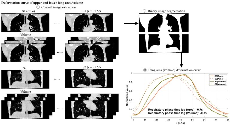
Fig. 2. Stitching methods, including time scale normalized cross-correlation analysis, time scale structural similarity analysis, sternum motion curve, deformation curve of upper and lower lung area/volume.
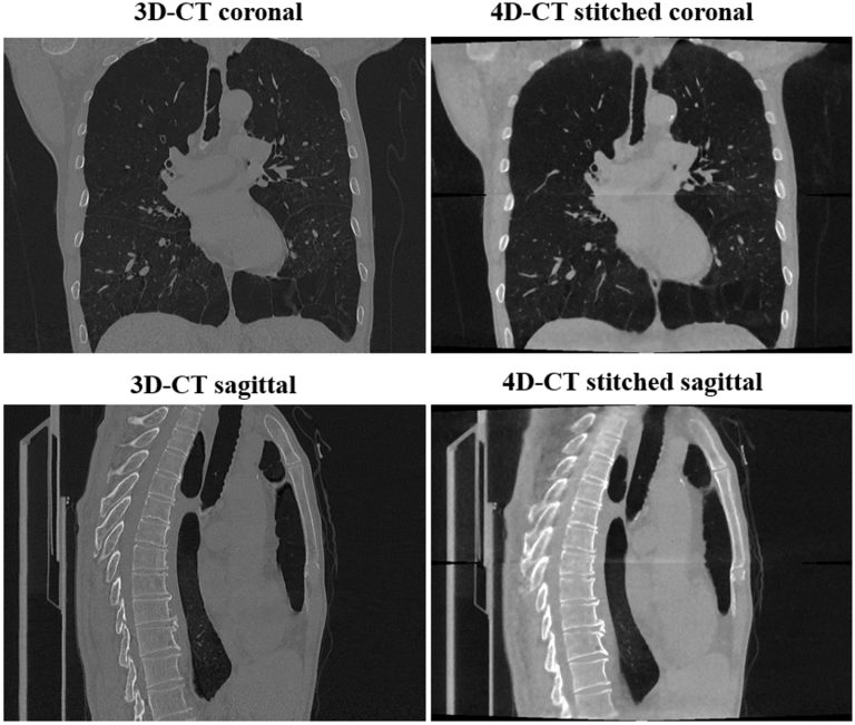
4D-CT stitching validation
The correlation value and comparison of the cross-sectional area of small pulmonary vessels between the stitched 4D-CT and 3D-CT breath-hold image.
Fig. 3. Comparison of the coronal and sagittal planes between the stitched 4D-CT and 3D-CT, with the normalized cross-correlation values.
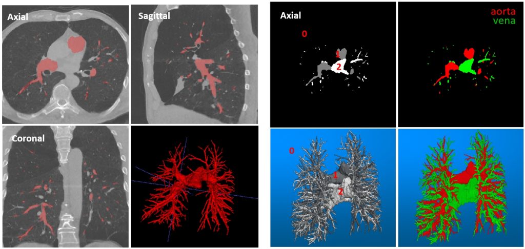
Fig. 4. Pulmonary vascular reconstruction and segmenting arteries and veins.